What is lung cancer staging, and why is it important?
Lung cancer staging is a way of describing where the lung cancer is located, if or where it has spread, and whether it is affecting other parts of the body. Treatment options are available for all stages of lung cancer, and knowing the stage helps your healthcare team:
- Understand how advanced your lung cancer is
- Recommend treatment options that are likely to be most effective for you
- Evaluate your response to treatment
There are differences in staging methods between the two major types of lung cancer—non-small cell lung cancer (NSCLC) and small cell lung cancer (SCLC).
NSCLC is staged by number as either I, II, III, or IV, where IV is the most advanced and indicates the cancer has spread. SCLC is typically classified as either early-stage or late-stage, but recently this type of cancer has been moving toward the same numbering system as NSCLC.
.png) To help you understand and share this information, you can request our free booklets on stages I, II, III, and IV non-small cell lung cancer that summarize the detailed information in the following sections.
To help you understand and share this information, you can request our free booklets on stages I, II, III, and IV non-small cell lung cancer that summarize the detailed information in the following sections.
When is lung cancer staged?
Lung cancer may be staged once or twice. The first staging, which all people should undergo, is carried out when someone is initially diagnosed and should be completed before treatment begins. This type of staging is called clinical staging. Clinical staging is based on the results of various tests, discussed in more detail in the Diagnosing Lung Cancer section, including imaging tests and biopsies. The clinical stage is not only the basis for deciding on a treatment plan, but is also the basis for comparison when checking into a person's response to treatment.
The second staging, called pathologic or surgical staging, adds what is learned about the individual's lung cancer from surgical treatment to the determination of staging. If the pathologic stage differs from the clinical stage (which it may, for example, if it is evident that the lung cancer has spread more than initially estimated), then the healthcare team can adjust the treatment more precisely.1,2
The TNM lung cancer staging system
The staging system described here is the system used for non-small cell lung cancer (NSCLC). including lung adenocarcinoma, squamous cell lung cancer, and large cell lung cancer. Stage classifications are based on the 8th edition American Joint Committee on Cancer lung cancer stage classifications.3,4,5,6,7,8
The TNM staging system can also be used for small cell lung cancer, although another staging system for small cell lung cancer that has only two stages, limited-stageCancer that is in the lung where it started and may have spread to the area between the lungs or to the lymph nodes above the collarbone and extensive-stageCancer that has spread widely throughout a lung, to the other lung, to lymph nodes on the other side of the chest, or to distant organs, is more often used.9
TNM stages are based on the values assigned to a patient's lung cancer in three categories—T (tumorAn abnormal mass of tissue that results when cells divide more than they should or do not die when they should), N (nodeLymph node—a rounded mass of lymphatic tissue surrounded by a capsule of connective tissue), and M (metastasisThe spread of cancer from the primary site, or place where it started, to other places in the body):
T: The size of the primary tumor and if it has grown into adjacent structures. The primary tumor is the original, or first, tumor
N: Whether and how regional lymph nodesA rounded mass of lymphatic tissue surrounded by a capsule of connective tissue are affected by the cancer. Lymph nodes are small bean-shaped structures that are part of the body's circulatory and immune systems: regional lymph nodes are those that are in the region around a tumor
M: Whether there is distant metastasis. Distant metastasis is the spread of cancer cells from the place where they first formed to distant organs, such as the adrenal glandA small gland that makes hormones; it helps control heart rate, blood pressure, and other important body functions. There is an adrenal gland on top of each kidney., bones, brain, or other lung, or to distant lymph nodes.
The particular combination of TNM values assigned to a patient's lung cancer determines its stage.
Stages using TNM classifications are designated by a number, zero (0) through four. For one through four, the Roman numerals I through IV are used. The lower the stage number, the less advanced the cancer is and the better the outcome is likely to be; the higher the stage number, the more advanced the cancer is. Stages I-IV are further divided into substages.
Stage 0
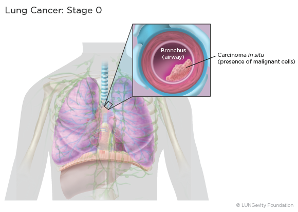
This is called "in situ" disease, meaning that the cancer is “in place” and has not spread from where it first developed.4,5,6,7,8,10,11
Stage I
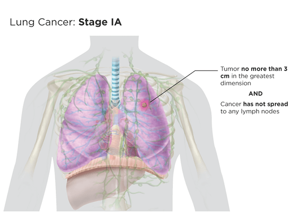
Stage I lung cancer tumors are small primary tumors that are in one lung only. Stage I lung cancer has not spread to any lymph nodes and has not metastasized. Stage I lung cancer is divided into two substages, stage IA and stage IB, based mainly on the size of the tumor.
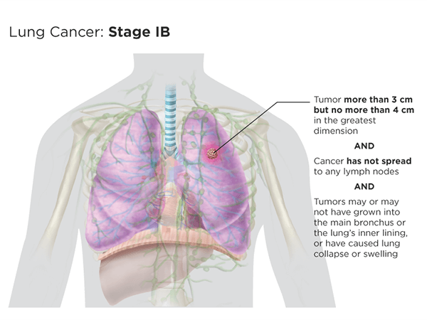
Smaller tumors, those no more than 3 centimeters (cm) in the greatest dimension, are stage IA, while slightly larger ones—more than 3 cm but no more than 4 cm in the greatest dimension—are stage IB. Stage IB tumors may or may not have grown into the main bronchus or the lung's inner lining or have caused lung collapse or swelling.4,5,6,7,8,10,11
Stage II
Like stage I lung cancer, stage II lung cancer is located in the lung where it has started. Stage II lung cancers have not metastasized to distant parts of the body.
Stage II lung cancer is divided into two stages: Stage IIA and stage IIB.4,5,6,7,8,10,11
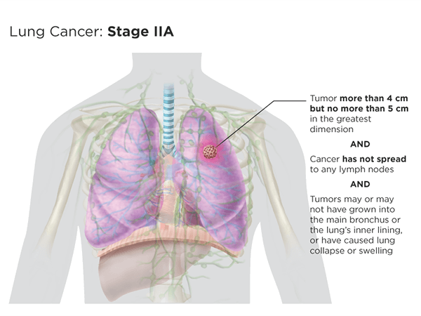
Stage IIA tumors are more than 4 cm but no more than 5 cm in the greatest dimension and have not spread to nearby lymph nodes. The tumors may or may not have grown into the main bronchus or into the lung's inner lining, or have caused lung collapse or swelling.
Stage IIB tumors are either:
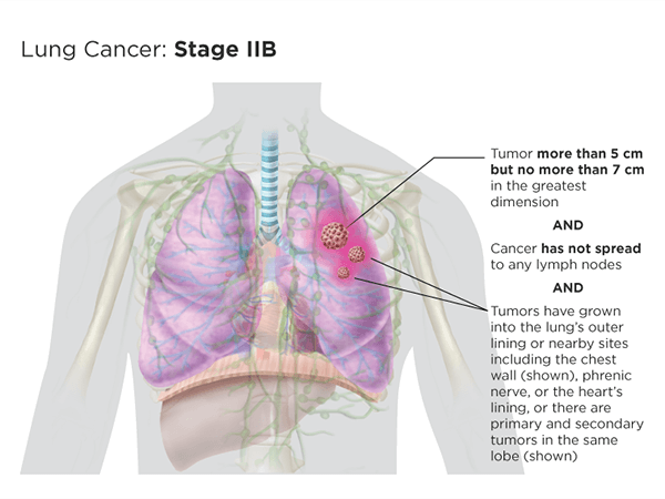
- more than 5 cm but no more than 7 cm in the greatest dimension and have not spread to nearby lymph nodes. They have grown into the lung's outer lining or nearby sites including the chest wall, phrenic nerveA nerve that runs from the spinal cord to the diaphragm (the thin muscle below the lungs and heart that separates the chest from the abdomen). It causes the diaphragm to contract and relax, which helps control breathing., or the heart's lining, or there are primary and secondary tumors in the same lobeA portion of an organ, such as the lung
OR
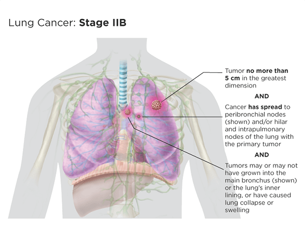
- no more than 5 cm in the greatest dimension and have spread to the peribronchialOf, relating to, occurring in, affecting, or being the tissues surrounding a bronchus nodes and/or to the hilarRelated to the hilum, on the medial side of each lung, where the main bronchus, pulmonary arteries, bronchial arteries, and nerves enter the lung and where the pulmonary veins, bronchial veins, and lymphatic vessels leave the lung and intrapulmonarySituated within, occurring within, or administered by entering the lungs nodes of the lung with the primary tumor. They may have grown into the main bronchus or into the lung's inner lining, or have caused lung collapse or swelling.4,5,6,7,8,10,11
Stage III
Stage III lung cancer has spread within the chest but has not metastasized to distant parts of the body. It is sometimes referred to as locally advanced.
Stage III lung cancer is divided into three stages: stage IIIA, stage IIIB, and stage IIIC.4,5,6,7,8,10,11
Stage IIIA tumors are either:
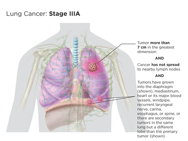
- more than 7 cm in the greatest dimension and have not spread to nearby lymph nodes. They may have grown into the diaphragmThe thin muscle below the lungs and heart that separates the chest from the abdomen, mediastinumThe area between the lungs. The organs in this area include the heart and its large blood vesels, the trachea, the esophagus, the thymus, and lymph nodes but not the lungs, heart or its major blood vessels, windpipe, recurrent laryngeal nerveThe branch of the vagus nerve supplying the larynx that arises below the larnyx, loops under the subclavian artery on the right side and under the arch of the aorta on the left, and returns upward to the larynx to supply all the muscles of the thyroid except the cricothyroid, carinaA ridge at the base of the trachea that separates the openings of the right and left main bronchi, esophagus, or spine, or there are secondary tumors in the same lung but a different lobe than the primary tumor
OR
- more than 5 cm in the greatest dimension and have spread to the peribronchial nodes and/or to the hilar and intrapulmonary nodes of the lung with the primary tumor. The tumors have grown into the lung's outer lining or nearby sites, including the chest wall, phrenic nerve, or the heart's lining, or there are primary or secondary tumors in the same lobe; and/or tumors have grown into the diaphragm, mediastinum, heart or its major blood vessels, windpipe, recurrent laryngeal nerve, carina, esophagus, or spine, or there are secondary tumors in the same lung but a different lobe than the primary tumor

OR
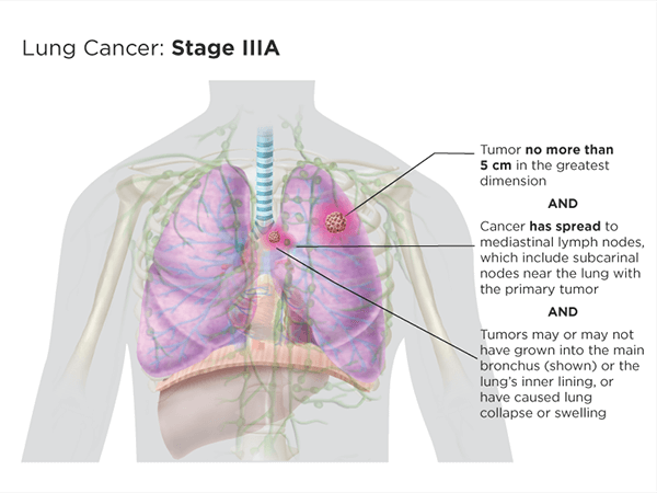
- no more than 5 cm in the greatest dimension and have spread to mediastinal lymph nodes, which include subcarinalLocated just below the carina of the trachea, where it splits into the right and left mainstem bronchi nodes, near the lung with the primary tumor. They may or may not have grown into the main bronchus or the lung's inner lining, or have caused lung collapse or swelling.
Stage IIIB tumors are either:
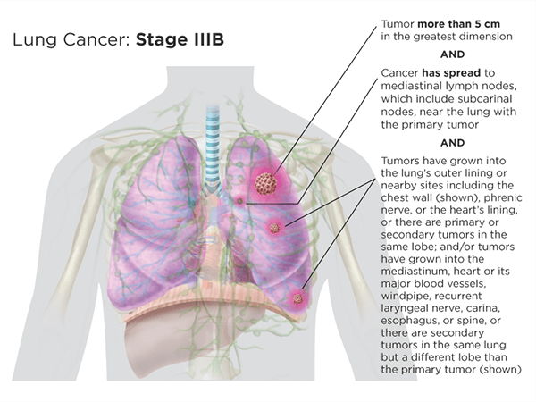
- more than 5 cm in the greatest dimension and have spread to mediastinal lymph nodes, which include subcarinal nodes, near the lung with the primary tumor. Tumors have grown into the lung's outer lining or nearby sites, including the chest wall, phrenic nerve, or the heart's lining, or there are primary or secondary tumors in the same lobe; and/or tumors have grown in the diaphragm, mediastinum, heart or its major blood vessels, windpipe, recurrent laryngeal nerve, carina, esophagus, or spine, or there are secondary tumors in the same lung but a different lobe than the primary tumor
OR
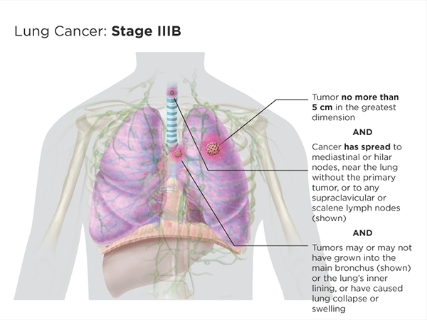
- no more than 5 cm in the greatest dimension and have spread to the mediastinal or hilar nodes near the lung without the primary tumor, or to any supraclavicularSituated or occurring above the clavicle, or scalene, lymph nodes. They may or may not have grown into the main bronchus or the lung's inner lining, or have caused lung collapse or swelling.
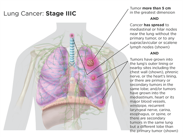
Stage IIIC tumors are more than 5 cm in the greatest dimension and have spread to mediastinal or hilar nodes near the lung without the primary tumor, or to any supraclavicular, or scalene, lymph nodes. Tumors have grown into the lung's outer lining or nearby sites, including the chest wall, phrenic nerve, or the heart's lining, or there are primary or secondary tumors in the same lobe, and/or tumors have grown into the diaphragm, mediastinum, heart or its major blood vessels, windpipe, recurent laryngeal nerve, carina, esophagus, or spine, or there are secondary tumors in the same lung but a different lobe than the primary tumor.
Stage IV
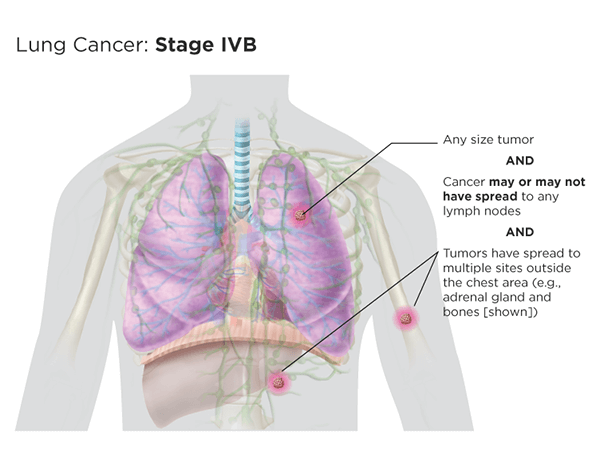
Unlike the earlier stages of lung cancer, stage IV lung cancer has metastasized to distant parts of the body. The tumors may be of any size and may or may not have spread to lymph nodes.
Stage IV lung cancer is divided into two stages: stage IVA and stage IVB.4,5,6,7,8,10,11
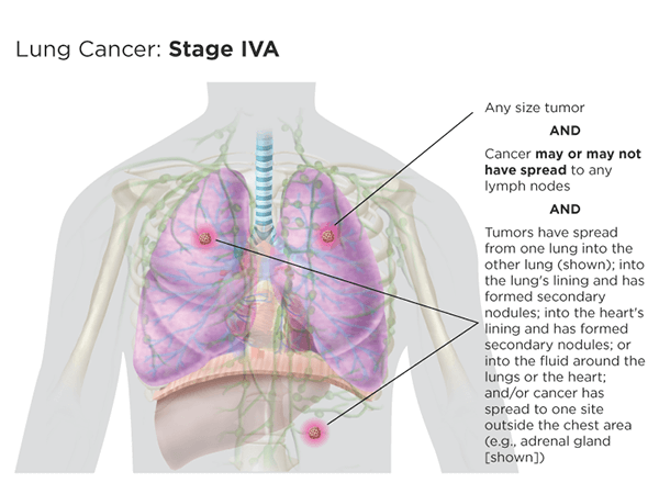
Stage IVA tumors may be of any size, may or may not have spread to any lymph nodes, and have metastasized, either from one lung into the other lung, into the lung's lining (and have formed secondary nodules), into the heart's lining (and have formed secondary nodules), or into the fluid around the lungs or the heart; and/or tumors have spead to one site outside the chest area (e.g., adrenal gland or bones).
Stage IVB tumors may be of any size, may or may not have spread to any lymph nodes, and have metastasized to multiple sites outside the chest area (e.g., adrenal gland and bones).4,5,6,7,8,10,11
Recurrence
Recurrent lung cancerLung cancer that has come back after a period of time during which the cancer could not be detected. The lung cancer may come back in the lung near the original tumor, in lymph nodes or in a distant organ. is lung cancer that has come back after treatment. If there is a recurrence, the cancer may need to be staged again ("restaged") using the system described above.8
Updated February 19, 2021
References
- Cancer Staging. American Cancer Society website. https://www.cancer.org/treatment/understanding-your-diagnosis/staging. Revised June 18, 2020. Accessed February 12, 2021.
- Cancer Staging. NIH National Cancer Institute website. https://www.cancer.gov/about-cancer/diagnosis-staging/staging. Posted March 25, 2015. Accessed Febreuary 12, 2021.
- Non-Small Cell Lung Cancer Stages. https://www.cancer.org/cancer/non-small-cell-lung-cancer/detection-diagnosis-staging/staging.html. Revised October 1, 2019. Accessed July 19, 2018.
- Lung Cancer—Non-Small Cell: Stages. Cancer.Net website. https://www.cancer.net/Cancer-types/lung-cancer-non-small-cell/stages. Approved May 2020. Accessed February 12, 2021.
- Detterbeck F, Boffa D, Kim A, Tanoue L. The Eighth Edition Lung Cancer Stage Classification. CHEST 2017; 151(1):193-203. https://journal.chestnet.org/article/S0012-3692(16)60780-8/fulltext. Accessed February 12, 2021.
- Kay FU, Kandathil A, Batra K, et al. Revisions to the tumor, node, metastasis staging of lung cancer (8th edition): rationale, radiologic findings and clinical implications. World Journal of Radiology. 2017 Jun 28:9(6):269-279. https://www.ncbi.nlm.nih.gov/pmc/articles/PMC5491654/. Accessed February 12, 2021.
- NCCN Guidelines for Patients®: Lung Cancer: Metastatic—Non-Small Cell Lung Cancer. Version 5.2019. https://www.nccn.org/patients/guidelines/content/PDF/lung-metastatic-patient.pdf. Posted June 7, 2019. Accessed February 12, 2021.
- Rami-Porta R, Asamura H, Travis W, Rusch V. Lung Cancer—major changes in the American Joint Committee on Cancer eigth edition cancer staging manual. CA: A Cancer Journal for Clinicians. Volume 67, Issue 2. March/April 2017. Pages 138-155. https://onlinelibrary.wiley.com/doi/10.3322/caac.21390/full. Accessed February 12, 2021.
- Small Cell Lung Cancer Stages. https://www.cancer.org/cancer/small-cell-lung-cancer/detection-diagnosis-staging/staging.html. Revised October 1, 2019. Accessed February 12, 2021.
- Detterbeck, FC. The eighth edition TNM stage classification for lung cancer: what does it mean on main street? Journal of Thoracic and Cardiovascular Surgery. Volume 155, Number 1. January 2018. https://www.jtcvs.org/article/S0022-5223(17)32136-0/pdf. Accessed February19, 2021.
- Non-Small Cell Lung Cancer Treatment (PDQ®) — Patient Version. NIH National Cancer Institute website. https://www.cancer.gov/types/lung/patient/non-small-cell-lung-treatment-pdq. Updated December 3, 2020. Accessed February 18, 2021.

